Department of Imaging Diagnostics and Interventional Radiology
The Department of Imaging Diagnostics and Interventional Radiology deals with the diagnosis of all neoplasms based on the analysis of the patient’s body in order to confirm or exclude the presence of pathologically changed tissues. The Department performs i.a. tests such as magnetic resonance imaging MRI, computed tomography CT, ultrasound, X-ray, mammography, etc.
MAGNETIC RESONANCE IMAGING
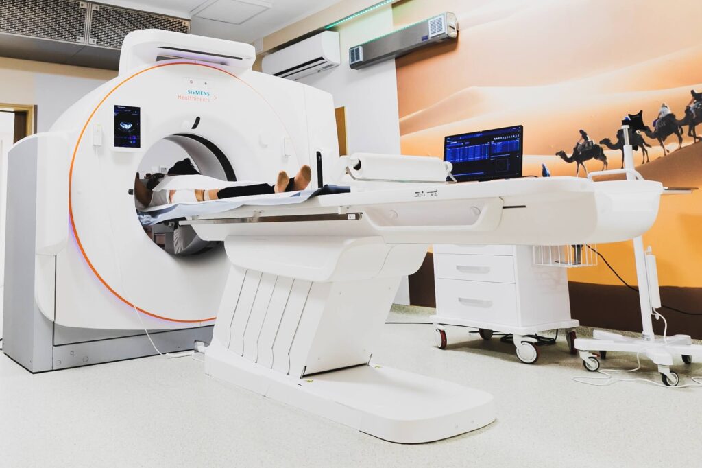
The Department of Imaging Diagnostics and Interventional Radiology has two MRI devices. One 1.5T and the second one latest generation 3T (three-Tesla) resonance. The Oncology Centre in Bydgoszcz is one of the few hospitals in the region that have such an apparatus. This modern device enables transabdominal prostate spectroscopy without the need to use a endorectal coil, brain spectroscopy is more accurate than with a 1.5T apparatus, it also allows to perform ASL perfusion without using a contrast agent.
Magnetic resonance imaging is an examination that uses a magnetic field produced by radio waves. It is non-invasive, painless and harmless, but requires the patient to remain motionless during the performance of individual sequences. The examination takes 15 to 60 minutes and is performed with or without contrast, depending on the medical indications.
Due to the better contrast resolution of the tissues, MRI examination allows to detect pathological changes, mainly in the brain, craniofacial region, pelvis, joints and bones more precisely than a CT scanner. We usually perform scans of the patient’s body in three sections – transverse, frontal and sagittal.
Obese or claustrophobic persons have great comfort during examinations because the magnetic resonance gantry is as wide as 70 cm.
For MR examination, it is necessary to estimate the level of creatinine in the blood or GFR (the examination is valid for 3 months). For patients with impaired kidney function, a contrast agent that is safe for them is administered or the examination is performed without a contrast agent (depending on the GFR level).
You should bring your previous test results (ultrasound, X-ray, CT, MRI, descriptions with photos or CD).
What should you inform your doctor about before MRI examination?
- about any metal implants in the body
- about having an installed pacemaker – absolute contraindication to the examination
- about claustrophobia
- about complications after previous MRI scans
- about pregnancy – the test is not performed in the first trimester of pregnancy
Preparation for the examination
- On the day of the examination, you should take medications that are taken continuously,
- The examination does not require any special preparation on the part of the patient, and there are no special restrictions on food consumption. Only in the case of examining the small intestine (enterography) and cholangiography MR, you should be on an empty stomach, take 1.5 litres of non-carbonated water with you, which you should start drinking two hours before the examination,
- If complications previously occurred after the administration of a contrast agent, it is advisable to administer prednisolone 30 mg (or methylprednisolone 32 mg) orally 12 hours and 2 hours before the administration of a contrast agent or to perform an examination without a contrast agent,
- If the patient is claustrophobic, the laboratory staff may offer him or her a mild sedative,
- When examining the head and eye sockets, patients are asked to forego makeup and hairspray, because cosmetic products contain metal particles that distort the results.
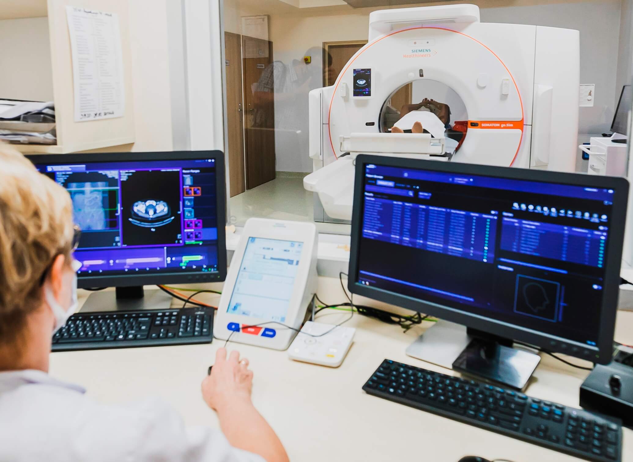
COMPUTER TOMOGRAPH (CT scanner)
Computed tomography is an examination with the use of ionizing radiation. The examination is painless, it takes 5-15 minutes, shows the internal organs and allows to detect any abnormalities.
Human organs differ in density, therefore they absorb different amounts of radiation. Due to this property, the patient’s tissues can be well differentiated. The images created by each rotation of the lamp are processed by the computer and presented on the monitor as an image of the patient’s body structures.
CT Scanner scans the patient’s body along one surface, but we can view and analyse it from different angles, most often in three sections – transverse, frontal and sagittal. CT is the examination of choice in the diagnosis of chest diseases.
Pregnancy is an absolute contraindication to CT examination. Therefore, every pregnant woman or a patient suspecting pregnancy should inform the medical staff of the laboratory about this fact. Exceptional cases are considered individually by the attending physician and carried out with the patient’s written consent.
For a CT scan, it is necessary to estimate the level of creatinine in the blood or GFR (the examination is valid for 3 months). For patients with impaired kidney function, a contrast agent that is safe for them is administered or the examination is performed without a contrast agent (depending on the GFR level).
You should bring your previous test results (ultrasound, X-ray, CT, MRI, descriptions with photos or CD).
Administration of contrast agents can induce
- dizziness,
- itching,
- feeling hot,
- nausea, vomiting,
- palpitations.
Any occurrence of the above should be reported to staff immediately.
Preparing for the examination
- it is recommended to drink 1l of non-carbonated water in the evening before the examination day and 2.5l after the examination,
- please arrive on an empty stomach or not eat and drink for 6 hours before the examination (patients with diabetes should take breakfast and medication with them),
- on the day of the examination, you should take medications that are taken constantly with a small amount of non-carbonated water (the exception is metformin)
- patients with unregulated hyperthyroidism or Graves-Basedov’s disease need the consent of an endocrinologist to administer a contrast agent.
X-RAY
X-ray examination is one of the basic diagnostic techniques in medicine. It is an examination with the use of ionizing radiation. We perform full-body examinations, except for images of teeth, as well as contrast examinations – urography, oesophagus examination and intestinal passage.
Ultrasound
It is an ultrasonic wave test. Ultrasound is a painless and harmless examination. We carry out examinations of all areas of the body, including Doppler, orthopaedic and rectal examinations.
For ultrasound examination of the abdominal cavity, please arrive on an empty stomach or 5-6 hours after eating, the urinary bladder should be full. Examination of other body areas does not require any preparation.
MAMMOGRAPHY
It is the basic examination in the diagnosis of breast cancer. It is an examination with the use of ionizing radiation. The painfulness of the examination depends on the individual sensitivity.
We perform diagnostic mammography from the age of 35 for medical indications, in symptomatic patients. We perform screening mammography in asymptomatic women between 50 and 69 years of age. These tests in our hospital are performed in the Department of Prevention and Health Promotion.
In over 95% of women, we do not find any changes suspected of being a malignant tumour. The remaining persons are called for tests as part of in-depth diagnostics: ultrasound, targeted and enlarged mammography images, fine- and large-needle biopsies, ultrasound-guided mammotomies and mammography (stereotaxic). We also perform galactography – an examination consisting in administering a contrast agent to the secreting milk duct and visualizing it in mammography. This examination is performed when intraductal hyperplasia is suspected.
 Kontakt do Zakładu

|
Coordinator of the Department of Imaging Diagnostics and Interventional Radiology
Jadwiga Szabo-Moskal, MD, PhD
|
|
Head of Medical Care
mgr Danuta Króliczewska
|